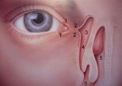
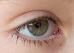
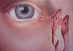
The lacrimal drainage system consists of the upper and lower lacrimal puncta, which continue as the lacrimal canaliculi. The canaliculi then open into the lacrimal sac, which drains tears into the nasolacrimal duct and subsequently into the nasal cavity.
The lacrimal drainage system drains tears and secretions from the eye into the nasal cavity. The various parts of the system can be affected by congenital malformations evident at birth, pathological processes or trauma. Disruption to drainage causes tears to accumulate in the conjunctival sac – a condition known as watering eyes or epiphora, and discharge occurs if infection is an issue.
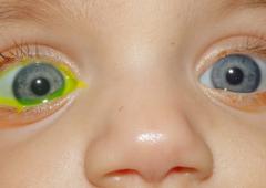
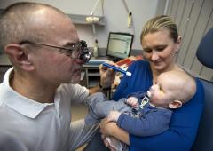
A relatively simple test can be carried out to evaluate the function of the lacrimal drainage system – the Fluorescein dye disappearance test (FDT). A drop of fluorescein is put into the conjunctival sac, after which the presence of any remaining dye is assessed. Drainage is normal if there is no dye (FDT grade 0). However, should any residual fluorescein remain in the conjunctival sac (FDT grade 1) or if the full dose is still present (FDT grade 2), then natural drainage is disrupted (Fig. 2). In such cases it is necessary to examine the lacrimal pathways to diagnose the problem. The FDT is an accurate test and even suitable for infants (Fig. 3).
Disorders can affect the areas of the canaliculi, lacrimal sac, nasolacrimal duct and nasal cavity.
Disorders of the Lacrimal Canaliculi
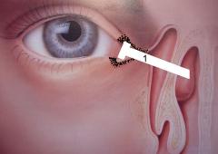
Most commonly, inflammation causes secretion from the lacrimal canaliculi and epiphora. The canaliculi are also prone to trauma. If a trauma or inflammation causes the canaliculi to narrow (stenosis) or block completely (obstruction) by up to 3 mm, it is possible to widen the stricture or remove the obstruction. This is done together with inserting a silicone cannula into the affected site for 3 months, a process referred to as stenting. In severe cases, if the unobstructed part of the canaliculi is at least 7 mm long, it is possible to remove the affected part surgically and re-attach the unobstructed part to the lacrimal sac, which opens into the nasal cavity. This procedure goes by the rather unwieldy name of canaliculodacryocystorhinostomy. Should the unobstructed part of the canaliculi be less than 7 mm long, it is necessary to bypass it and create an artificial drainage pathway between the conjunctival sac and the nasal cavity. The procedure to do this, conjunctivodacryocystorhinostomy (CDCR), involves inserting a special glass cannula called a Jones tube (Figs. 4, 5). It is necessary to clean the tube regularly, something that young children find challenging. Therefore, CDCR is only performed on patients over 10 years of age.
Disorders of the Lacrimal Sac and Nasolacrimal Duct
Disorders affecting the lacrimal sac are not very frequent, with acute or chronic inflammation (dacryocystitis) or formation of a tear stone (dacryolithiasis) being the usual issues encountered. However, disorders of the nasolacrimal duct are relatively common. Children often experience obstruction that relates to a congenital condition, i.e one existing at birth, known as congenital lacrimal obstruction (see Congenital Lacrimal Obstruction). In adults, such an obstruction is usually acquired through inflammation or trauma. The obstruction is either treated by stenting the nasolacrimal duct with a silicone cannula, or by creating an incised opening (stoma) between the lacrimal sac and nasal cavity (Fig. 6). This procedure is called dacryocystorhinostomy (DCR), and can be performed from the side of the face (external DCR) or via the nasal cavity (endonasal DCR, EDCR).
Our Clinic carries out unique diagnostic procedures to treat the lacrimal drainage system, and over 12,000 adult and pediatric patients – from the Czech Republic and abroad – have received treatment over the years.
